Cat. No Product Name Pack Size
LF-SAB5001 mouse/rabbit IgG SABC Kit (HRP) 1kit
LF-SAB5002 mouse IgG SABC Kit (HRP) 1kit
LF-SAB5003 rabbit IgG SABC Kit (HRP) 1kit
LF-SAB5004 goat IgG SABC Kit (HRP) 1kit
LF-SAB5005 human IgG SABC Kit (HRP) 1kit
LF-SAB5006 rat IgG SABC Kit (HRP) 1kit
LF-SAB5007 mouse/rabbit IgG SABC Kit (HRP), Ready to use 1kit
LF-SAB5008 mouse IgG SABC Kit (HRP), Ready to use 1kit
LF-SAB5009 rabbit IgG SABC Kit (HRP), Ready to use 1kit
LF-SAB5010 goat IgG SABC Kit (HRP), Ready to use 1kit
LF-SAB5011 human IgG SABC Kit (HRP), Ready to use 1kit
LF-SAB5012 rat IgG SABC Kit (HRP), Ready to use 1kit
LF-SAB5013 mouse IgM SABC Kit (HRP), Ready to use 1kit
LF-SAB5014 mouse IgG SABC Kit (HRP), Ready to use 1kit
LF-SAB5015 rabbit IgG SABC Kit (HRP), Ready to use 1kit
LF-SAB5016 mouse/rabbit IgG SABC Kit (AP), Ready to use 1kit
LF-SAB5017 mouse IgG SABC Kit (AP), Ready to use 1kit
LF-SAB5018 rabbit IgG SABC Kit (AP), Ready to use 1kit
LF-SAB5019 goat IgG SABC Kit (AP), Ready to use 1kit
LF-SAB5020 human IgG SABC Kit (AP), Ready to use 1kit
LF-SAB5021 rat IgG SABC Kit (AP), Ready to use 1kit
LF-SAB5022 mouse IgM SABC Kit(AP), Ready to use 1kit
LF-SAB5023 mouse IgG SABC Kit (FITC) 1kit
LF-SAB5024 mouse IgM SABC Kit (FITC) 1kit
LF-SAB5025 rabbit IgG SABC Kit (FITC) 1kit
LF-SAB5026 goat IgG SABC Kit (FITC) 1kit
LF-SAB5027 human IgG SABC Kit (FITC) 1kit
LF-SAB5028 rat IgG SABC Kit (FITC) 1kit
LF-SAB5029 mouse IgG SABC Kit (FITC+POD) 1kit
LF-SAB5030 mouse IgM SABC Kit (FITC+POD) 1kit
LF-SAB5031 rabbit IgG SABC Kit (FITC+POD) 1kit
LF-SAB5032 goat IgG SABC Kit (FITC+POD) 1kit
LF-SAB5033 human IgG SABC Kit (FITC+POD) 1kit
LF-SAB5034 rat IgG SABC Kit (FITC+POD) 1kit

SABC kit protocol
- Streptavidin is a 62,000 molecular weight with an extraordinarily high affinity for the small molecular weight vitamin, biotin.
- Because this affinity is over one million times higher than that of antibody for most antigens, the binding of Streptavidin to biotin (unlike antibody-antigen interactions) is essentially irreversible.
- In addition to this high affinity, the Biotin/ Streptavidin System can be effectively exploited because Streptavidin has four binding sites for biotin and most proteins (including antibodies and enzymes) can be conjugated with several molecules of biotin. These aspects provide the potential for macromolecular complexes to be formed between Streptavidin and biotinylated enzymes.
- An immunoperoxidase procedure based on these properties was devised for localizing a variety of histologically significant antigens and other markers. (Hsu SM, Raine L, Fanger H: Am. J. Clin. Pathol. 7 5, 734-738, 1981; Hsu SM, Raine L, Fanger H: J. Histochem. Cytochem. 2 9, 577-580, 1981) this technique employs unlabeled primary antibody, followed by biotinylated secondary antibody and then a preformed Avidin and Biotinylated horseradish peroxidase macromolecular Complex.
- Abfrontier SABC kit is with Streptavidin and biotinylated horseradish peroxidase reagents, which have been specially prepared to form ideal complexes for immunoperoxidase staining. Although the structure of the Streptavidin: biotinylated horseradish peroxidase complex is still undefined, evidence suggests that it consists of many biotinylated horseradish peroxidase molecules crosslinked by Streptavidin into a three dimensional array.
- The complex apparently has few exposed biotin residues but retains at least one biotin binding site. Formation of the complex is achieved by mixing Streptavidin and biotinylated horseradish peroxidase in dilute solution and in defined amounts prior to use.
- After the initial incubation there appears to be little change in the complex as judged by only a marginal increase in immune peroxidase staining sensitivity and the complex remains stable for several hours after formation. The high sensitivity and shorter incubation times reported for the best SABC system are likely due to the number of active horseradish peroxidase molecules associated with the complex and the rapid, irreversible interaction of the complex with biotinylated antibody.
- The low background staining obtainable with the Abfrontier SABC Kits is probably due to the high dilutions of primary antisera and other reagents employed in the method, the quality of our affinity-purified biotinylated secondary antibodies, and the specially prepared Streptavidin and biotinylated horseradish peroxidase.

| LF-SAB5003 | rabbit IgG SABC Kit (HRP) | 1kit | Abfrontier |
|
|
|
|||
| LF-SAB5014 | mouse IgG SABC Kit (HRP), Ready to use | 1kit | Abfrontier |
|
|
|
|||
| LF-SAB5033 | human IgG SABC Kit (FITC+POD) | 1kit | Abfrontier |
|
|
|
|||
| LF-SAB5016 | mouse/rabbit IgG SABC Kit (AP), Ready to use | 1kit | Abfrontier |
|
|
|
|||
| LF-SAB5020 | human IgG SABC Kit (AP), Ready to use | 1kit | Abfrontier |
|
|
|
|||
| LF-SAB5012 | rat IgG SABC Kit (HRP), Ready to use | 1kit | Abfrontier |
|
|
|
|||
| LF-SAB5010 | goat IgG SABC Kit (HRP), Ready to use | 1kit | Abfrontier |
|
|
|
|||
| LF-SAB5023 | mouse IgG SABC Kit (FITC) | 1kit | Abfrontier |
|
|
|
|||
| LF-SAB5013 | mouse IgM SABC Kit (HRP), Ready to use | 1kit | Abfrontier |
|
|
|
|||
| LF-SAB5017 | mouse IgG SABC Kit (AP), Ready to use | 1kit | Abfrontier |
|
|
|
|||
| LF-SAB5030 | mouse IgM SABC Kit (FITC+POD) | 1kit | Abfrontier |
|
|
|
|||
| LF-SAB5027 | human IgG SABC Kit (FITC) | 1kit | Abfrontier |
|
|
|
|||
| LF-SAB5019 | goat IgG SABC Kit (AP), Ready to use | 1kit | Abfrontier |
|
|
|
|||
| LF-SAB5005 | human IgG SABC Kit (HRP) | 1kit | Abfrontier |
|
|
|
|||
| LF-SAB5021 | rat IgG SABC Kit (AP), Ready to use | 1kit | Abfrontier |
|
|
|
|||
| LF-SAB5002 | mouse IgG SABC Kit (HRP) | 1kit | Abfrontier |
|
|
|
|||
| LF-SAB5008 | mouse IgG SABC Kit (HRP), Ready to use | 1kit | Abfrontier |
|
|
|
|||
| LF-SAB5009 | rabbit IgG SABC Kit (HRP), Ready to use | 1kit | Abfrontier |
|
|
|
|||
| LF-SAB5032 | goat IgG SABC Kit (FITC+POD) | 1kit | Abfrontier |
|
|
|
|||
| LF-SAB5028 | rat IgG SABC Kit (FITC) | 1kit | Abfrontier |
|
|
|
|||
| LF-SAB5024 | mouse IgM SABC Kit (FITC) | 1kit | Abfrontier |
|
|
|
|||
| LF-SAB5022 | mouse IgM SABC Kit(AP), Ready to use | 1kit | Abfrontier |
|
|
|
|||
| LF-SAB5018 | rabbit IgG SABC Kit (AP), Ready to use | 1kit | Abfrontier |
|
|
|
|||
| LF-SAB5007 | mouse/rabbit IgG SABC Kit (HRP), Ready to use | 1kit | Abfrontier |
|
|
|
|||
| LF-SAB5034 | rat IgG SABC Kit (FITC+POD) | 1kit | Abfrontier |
|
|
|
|||
| LF-SAB5006 | rat IgG SABC Kit (HRP) | 1kit | Abfrontier |
|
|
|
|||
| LF-SAB5029 | mouse IgG SABC Kit (FITC+POD) | 1kit | Abfrontier |
|
|
|
|||
| LF-SAB5025 | rabbit IgG SABC Kit (FITC) | 1kit | Abfrontier |
|
|
|
|||
| LF-SAB5026 | goat IgG SABC Kit (FITC) | 1kit | Abfrontier |
|
|
|
|||
| LF-SAB5001 | mouse/rabbit IgG SABC Kit (HRP) | 1kit | Abfrontier |
|
|
|
|||
| LF-SAB5015 | rabbit IgG SABC Kit (HRP), Ready to use | 1kit | Abfrontier |
|
|
|
|||
| LF-SAB5031 | rabbit IgG SABC Kit (FITC+POD) | 1kit | Abfrontier |
|
|
|
|||
| LF-SAB5004 | goat IgG SABC Kit (HRP) | 1kit | Abfrontier |
|
|
|
|||
| LF-SAB5011 | human IgG SABC Kit (HRP), Ready to use | 1kit | Abfrontier |
|
|
|
|||
| SA1054 | Alkaline Phosphatase Conjugated anti-Human IgG SABC Kit | 1 kit | Boster | IHC-P;IHC-F;ICC |
|
|
|
||
| SA1028 | HRP Conjugated anti-Rabbit IgG SABC Kit | 1 kit | Boster | IHC-F;ICC |
|
|
|
||
| SA1050 | Alkaline Phosphatase Conjugated anti-Mouse/Rabbit IgG SABC Kit | 1 kit | Boster | IHC-P;IHC-F;ICC |
|
|
|
||
| SA1056 | Alkaline Phosphatase Conjugated anti-Mouse IgM SABC Kit | 1 kit | Boster | IHC-P;IHC-F;ICC |
|
|
|
||
| SA1082 | FITC + POD Conjugated anti-Rabbit IgG SABC Kit | 1 kit | Boster | IHC-P;IHC-F;ICC |
|
|
|
||
| SA1093 | DyLight®488 Conjugated anti-Mouse IgM SABC Kit | 1 kit | Boster | IHC-P;IHC-F;ICC |
|
|
|
||
| SA1092 | DyLight®488 Conjugated anti-Mouse IgG SABC Kit | 1 kit | Boster | IHC-P;IHC-F;ICC |
|
|
|
||
| SA1099 | DyLight®488 Conjugated anti-Rat IgG SABC Kit | 1 kit | Boster | IHC-P;IHC-F;ICC |
|
|
|
||
| SA1038 | HRP Conjugated Donkey Anti-Rat IgG SABC Kit No Cross-Reactivity With Mouse Serum Protein | 1 kit | Boster | IHC-P;IHC-F;ICC |
|
|
|
||
| SA1094 | DyLight®488 Conjugated anti-Rabbit IgG SABC Kit | 1 kit | Boster | IHC-P;IHC-F;ICC |
|
|
|
||
| SA1074 | Cy3 Conjugated anti-Rabbit IgG SABC Kit | 1 kit | Boster | IHC-P;IHC-F;ICC |
|
|
|
||
| SA1051 | Alkaline Phosphatase Conjugated anti-Mouse IgG SABC Kit | 1 kit | Boster | IHC-P;IHC-F;ICC |
|
|
|
||
| SA1025 | HRP Conjugated anti-Rat IgG SABC Kit | 1 kit | Boster | IHC-P;IHC-F;ICC |
|
|
|
||
| SA1021 | HRP Conjugated anti-Mouse IgG SABC Kit | 1 kit | Boster | IHC-P;IHC-F;ICC |
|
|
|
||
| SA1024 | HRP Conjugated anti-Human IgG SABC Kit | 1 kit | Boster | IHC-P;IHC-F;ICC |


INSTRUCTIONS FOR IMMUNOHISTOCHEMICAL STAINING INTRODUCTION
Reagents supplied (for 250 sections)
component size storage
Blocking Serum 20ml*2 Store at 4°C for 1year
Biotinylated,Affinity-purified Anti-Rabbit IgG 500ul Store at 4°C for 1year
Reagent A (Streptavidin) 500ul Store at 4°C for 1year
Reagent B (Biotinylated Horseradish Peroxidase) 500ul Store at 4°C for 1year
NOTE: reagents not supplied (Primary Antibody, Buffer, Hydrogen Peroxide, Oxidizable Peroxidase Substrate)
PREPARATION OF WORKING SOLUTIONS
A number of different buffers can be used in the Abfrontier SABC system. One of the most common is 10 mM sodium phosphate, pH 7.4, 0.9% saIine (PBS). The SABC working solutions are prepared as follows:
Blocking Serum working solution: add 2ml of stock Blocking Serum to 10 ml of PBS. The preferred serum for blocking is prepared from the same species in which the biotinylated secondary antibody is made.
Biotinylated Antibody working solution: add 100ul Biotinylated Antibody to 10 ml of Blocking Serum working solution (PBS contain Blocking Serum).
SABC Reagent working solution: Mix 100ul of Reagent A and 100ul Reagent B in a EP tube, immediately. Then place it on 37°C for 30 Minutes, add the mixture into 10 ml of Blocking Serum working solution(PBS contain Blocking Serum)for using. It is best to use SABC Reagent working solution within 12 hours.
The configuration table
Blocking Serum Working solution Biotinylated Antibody Working solution SABC Reagent Working solution
1 section 200ul-400ul 200ul 200ul
10 sections 2ml-4ml 2ml 2ml
100 sections 20ml-40ml 20ml 20ml
250 sections 50ml-100ml 50ml 50ml
STAINING PROCEDURE FOR PARAFFIN SECTIONS
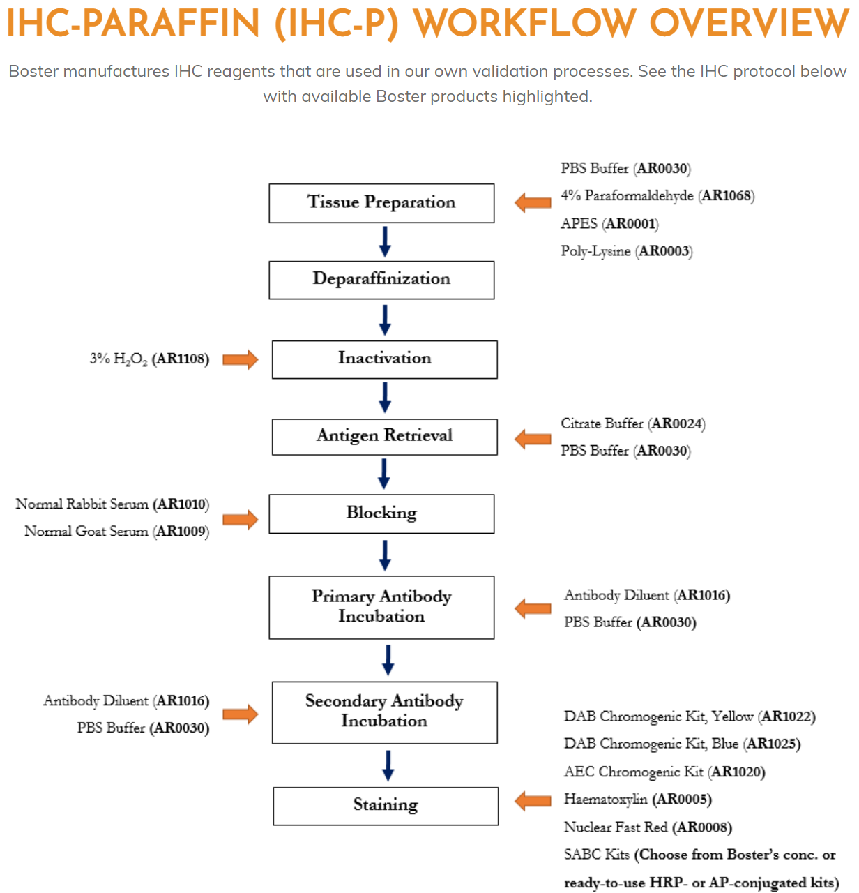
Deparaffinize and hydrate tissue sections through xylenes or other clearing agents and graded alcohol series.
Rinse for 5 minutes in tap water.
If quenching of endogenous peroxidase activity is required, incubate the sections for 30 minutes in 0.3% H2O2 in methanol or water. Incubation times may be shortened by using higher concentrations of H2O2. If endogenous peroxidase activity does not present a problem, step 3 may be deleted.
Wash in buffer for 5 minutes.
Incubate sections for 60 minutes with diluted Blocking Serum working solution from the species in which the secondary antibody is made. (In cases where non-specific staining is not a problem, Steps 5 and 6 may be deleted).
Blot excess Blocking Serum working solution from sections.
Incubate sections for 60 minutes with primary antiserum diluted in buffer. (If background staining occurs, dilutions of the primary and secondary antibodies may be made in buffer containing 1-2% of the appropriate blocking serum.)
Wash slides for 5 minutes in buffer.
Incubate sections for 60 minutes with diluted biotinylated secondary antibody solution.
Wash slides for 5 minutes in buffer.
Incubate sections for 30 minutes with Abfrontier SABC Reagent working solution
Wash slides for 5 minutes in buffer.
Incubate sections in peroxidase substrate solution until desired stain intensity develops. (See Note 1)
Rinse sections in tap water. 15. Counterstain, clear and mount.
NOTES
Solutions containing sodium azide or other inhibitors of peroxidase activity should not be used in diluting the peroxidase substrate or the SABC Reagent. Do not add normal serum, non-fat dried milk, culture media, or other potential sources of biotin to the SABC reagent. This may result in reduced sensitivity.
Development times may differ depending upon the level of antigen, the intensity of the stain that is required, or the substrate used. DAB generally should be developed for 2-10 minutes; AEC for 10-30 minutes; TMB for 5-20 minutes. Some counterstains may not be compatible with certain peroxidase substrates because of solubility of the reaction products or lack of color contrast. A counterstain compatibility chart is available upon request.
If the reagents are to be diluted beyond their recommended concentrations, first prepare the diluted biotinylated antibody and SABC reagent as described in the instructions. Subsequent dilutions should be made in a buffer containing 0.1% immunohistochemical grade bovine serum albumin. Only immunohistochemical grade BSA should be used, as other preparations can contain undesired impurities. Dilution of these reagents may require longer incubation times and/or higher incubation temperatures to achieve maximum sensitivities.
The section should be well prepared. Fixation (generally, in buffered formalin not exceeding 4 percent formaldehyde) should be sufficient to maintain the integrity of the section throughout the staining procedure but not so harsh as to destroy the antigen under study. During the staining procedure, do not allow the section to dry out. Use a humidified chamber for incubations.
To avoid adsorption of the antibody to the plastic or glass container in which the final dilution is made, the primary antibody may be diluted in buffers containing 0.1% immunohistochemical grade bovine serum albumin or dilute Blocking Serum.
Incubation times may be shortened. In cases where the antigen concentration in the section is high, suggested incubation times with primary antibody, biotinylated secondary antibody, and SABC Reagent may be reduced. Incubation times as short as five minutes have been reported to be sufficient in some cases when incubation temperatures are raised to 37 °C. If the antigen concentration is low, steps 7 and 9 may be lengthened to achieve maximal staining